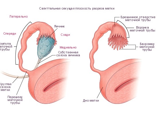
Structure of the Fallopian tube
Fallopian tubes consist of four sections throughout their entire length. They move away from the body of the uterus almost horizontally and end in an enlarged fringe part, which has the name of a funnel. These are the widest parts of the tube in the immediate vicinity of the ovary, in which an egg is born and comes out on a certain day of the menstrual cycle to meet the sperm.
Further, after the funnel, there is an ampular section of the tube - a fairly wide part of it. After this, the uterine or fallopian tube gradually narrows, and this part of the isthmus is called isthmic.
The tubes end in the uterine part, where they pass into this muscular organ. The walls of the pipes differ in their structure: the outer layer is a serous membrane (the peritoneum), the middle one consists of the longitudinal and circular layer of muscles, and the inner layer is the mucosa, collected in the grooves and covered with the ciliated epithelium, by means of which the egg moves to the uterine cavity.
Size of the fallopian tube
Fallopian tubes, in spite of their important function, have very small dimensions. The length of one is from 10 to 12 cm, and the width (or rather, the diameter) is only 0.5 cm. If a woman has any disease of the fallopian tubes, then a slight increase in diameter is possible, due to edema or inflammation.
Function of the fallopian tubes
On one of the segments of the path, the egg under favorable conditions meets with a sperm and conception occurs, that is, the birth of a new life. Further, thanks to the lining of the inner villous epithelium, the fertilized egg moves into the uterine cavity, where after a lapse of 5-7 days the pathway is implanted into its muscular layer. So begins the pregnancy, which will last 40 weeks.