Sometimes in ultrasound-findings in the ovary - left or right the doctor writes about the presence of anechogenous formations. Echogenicity is the term used in ultrasound diagnostics to indicate the conductivity of ultrasonic waves by tissues. Such tissues as bone completely reflect ultrasound because of its high density, and it is completely reflected on the border of organs and tissues that contain air. Thick fabrics reflect ultrasound more strongly, and those that contain a lot of liquid conduct the signal of the ultrasonic sensor, strengthening it at the same time.
The ultrasound signal and dense tissues (bone) reflected from organs and tissues are reflected on the screen of the monitor, and the air will look white (hyperechoic), the signal does not pass after them, and behind them there is a black band equal to the reflected signal (acoustic shadow). The more dense weave, the higher its echogenicity (the lighter it looks), the more water contains tissue or organ (including blood vessels with blood) - the lower its echogenicity, and the fluid formations will be anechogenous (black).
Structure of the ovary on ultrasound
Often there is an anechoic cavity of various sizes inside the ovary. To understand what a normal ovary and an anechoic ovarian cyst look like on ultrasound, you should know what changes occur in the normal menstrual cycle. After the end of menstruation, follicles begin to grow in one or both ovaries: a small anehogenous inclusion of a circular shape in the ovary with a size of 1-3 mm grows to 7-8 mm, this occurs in the first half of the cycle. Then one, from follicles becomes dominant - it continues to grow in sizes from 16-17 to 25-30 mm, from it during ovulation the egg leaves.
After the release of the ovule, the circular anehogenous formation slightly diminishes in size, becomes irregular in shape, turning into a yellow body. 2-3 days before the onset of menstruation, the yellow body stops working and often bursts, releasing a small amount of fluid, therefore, from the beginning and until the end of menstruation in the ovaries there should be no anechogenic formations.
If pregnancy has occurred, then the yellow body functions the first trimester of pregnancy and looks like an anechogenous formation of a circular shape on one of the ovaries (the yellow body of pregnancy that produces progesterone).
Ovarian cysts on ultrasound
Various disorders of the hormonal background in a woman and the function of her ovaries can lead to the appearance of other anechogenous formations - ovarian cysts.
- Most often on one of the ovaries, a follicular cyst is found - anechogenous formation of a round form, of a homogeneous structure with a thin capsule, measuring from 3 to 6 cm in diameter. It occurs with hormonal disorders that lead to the absence of ovulation - the egg does not leave the follicle, which continues to grow in size. Follicular cysts themselves disappear during 1-3 menstrual cycles, less often, complicated, they require appropriate treatment.
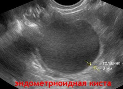
- Often on the ovaries another anechogenous formation is found - the endometrioid cyst . A distinctive feature of this formation is a harder capsule,
heterogeneity of the cyst and its constant size or growth over many menstrual cycles. The size of the endometrioid cyst can be different - from a few millimeters to several centimeters, cysts with endometriosis are single and multiple. - Other anehogennye formations - single or multi-chambered serous cysts, can not only be an independent entity, but also the manifestation of another, for example, a malignant tumor. Multichamber, heterogeneous echopositive inclusions or proliferation on the walls inside such anechogenous structures may indicate a malignant process in the ovaries.