If symmetry, size, shape and density of the mammary glands are not affected, as well as unpleasant and painful sensations in one or both of the mammary glands, every woman should undergo an ultrasound examination and a consultation with a mammologist as soon as possible.
Fibrous-cystic formations in the mammary gland, visible on the apparatus of ultrasound
Echogenicity of the mammary gland is determined by the degree of density (cellularity) of tissues and their different vision on the monitor of the ultrasound apparatus.
- Anehogenous formation in the mammary gland is a cyst that is diagnosed when examining the ultrasound of the mammary glands , and the danger of this disease is detected by puncture and cytological examination of its contents.
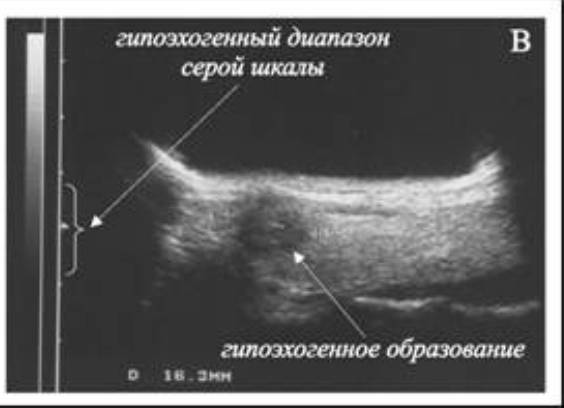
- Often in middle-aged women, hormone changes may reveal a hypoechoic formation of the breast, it can be a benign tumor or cystic formation. As a rule, hypoechoic formations turn out to be a fluid accumulation, especially if its size does not exceed 1 cm. If the formation increases, a biopsy should be done for histological examination.
Echogenic formations in the mammary gland are knotty seals with dense walls and liquid contents. As in the above processes, a more detailed study using biopsy and content analysis is needed to clearly define the content and the purpose of the appropriate treatment.
- Isoechoic formation of the mammary gland. This type of nodal benign breast tumors corresponds to normofollicular adenoma.
- Hyperechoic formation in the mammary gland is a rather large compaction of the echogenic structure.
- Hypoechoic avascular formation of the mammary gland has a more complex structure and can have both fluid and vascular elements of various sizes. The formation of reduced echogenicity of the breast is characterized by a weak reflection on the ultrasound monitor, this factor is more characteristic of tumor formations, but it also happens in cystic breast tumors .