Uterus in its structure is a unique organ, the most important in the reproductive system of a woman. In connection with the increased incidence of genital organs and sometimes inability to get help from a qualified specialist, every woman must be familiar with the structure and functions of the uterus.
The structure of the womb is a general characteristic
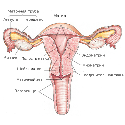
The uterus is a smooth-muscular hollow organ, the main function of which is aimed at bearing the fetus and its subsequent expulsion. It consists of three parts:
- Cervix of the uterus . This muscular ring that connects the uterus to the vagina performs a protective function. Inside the cervix is an opening, the so-called cervical canal, its glands produce mucus, which prevents the penetration of pathogenic bacteria into the uterine cavity.
- Isthmus - the transition between the neck and the body of the uterus, the main function is to open and exit the fetus.
- The main body is the basis of the whole organ, the place of origin and development of a new life.
The size of the uterus varies depending on the age of the woman, the number of births and pregnancies. Thus, in a nulliparous woman its length is 7-8 cm, width - 5 cm, weight does not exceed 50 g. After repeated reproduction of the offspring, the size and weight increase. Due to the peculiarities of the structure, during pregnancy the uterus can stretch up to 32 cm in length and up to 20 cm in width. These abilities are laid down at the genetic level and are activated under the influence of the hormonal background. The main principles of the structure of the uterus are aimed at creating favorable conditions for the development of the fetus during pregnancy.
Histological structure of the uterus
The structure of the uterine wall is three-layered and has no other analogues.
- The first inner layer is the mucous membrane , in medical practice is called the endometrium . Contains a large number of blood vessels and is subject to cyclic changes. All processes in the endometrium are directed to the embryo; if pregnancy does not occur, its surface layer is rejected, in fact this is menstruation. The structure and functions of the uterus, namely, its mucous membrane during pregnancy, can provide nutrients and create comfortable conditions for the life of the fetus.
- The second layer is smooth muscle fibers , interwoven in all directions, called the myometrium. Have the property of shrinking. In the normal state, the myometrium shrinks during sexual intercourse or menstruation. In pregnancy, despite its structure, the female organism is blocked as much as possible this feature, that is, for a favorable bearing the uterus should be relaxed. By the time of birth, the myometrium significantly increases in size, thereby allowing its fetuses to expel the fetus.
- The third layer is the perimeter . It is a connective tissue that connects the uterus to the peritoneum. At the same time it leaves the necessary minimum for movements in case of any changes in neighboring organs.
Diseases of the uterus
Most often, problems with the functionality of this organ are manifested in the form of menstrual
As consequences, miscarriage, infertility, inflammation and other unpleasant moments may develop.
Summing up, we can conclude that the structure of the uterus and appendages in the female body is aimed at the reproduction of a new life. All the changes that occur in this body are controlled by hormones and other biologically active substances. If a woman has not been previously or in the process of pregnancy, any diseases of the genitourinary system, other organs, infection of various etiologies, including venereal, it can be confidently said that nature will take care of the safe birth of a healthy baby.